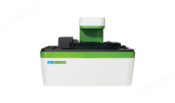
MuviCyte Live-Cell Imaging Kit活细胞成像系统
产品简介
详细信息
The MuviCyte live-cell imaging system is designed to operate inside your cell-culture incubator, enabling you to maintain your cells under optimal conditions and perform a wide range of assays in a variety of culture vessels.
Life science research labs study cellular behaviors and pathways to gain a deeper understanding of functions, disease mechanisms and responses to treatments. Live-cell imaging is a vital tool for getting maximum information from precious cell samples.
The MuviCyte live-cell imaging kit consists of a MuviCyte live-cell imaging instrument plus 4x, 10x and 20x objective lenses, PC and monitor
The MuviCyte system is designed to operate inside your incubator, so you can maintain your cells under optimal conditions, keeping them healthy for weeks at a time. Controlled by an external PC, you can observe your cells remotely, which helps to keep the culture chamber at optimum levels of temperature, CO2, and humidity. The automated operation allows you to focus on your science while the instrument runs unattended.
With three-color fluorescence imaging, z-stacking, and stitching capabilities, you can perform a wide range of assays in a variety of culture vessels, e.g. chamber slides, petri dishes, flasks, microplates. Automated imaging, over hours to days, means your assays can be run at much higher throughput than with a traditional microscope.
The system has built-in image analysis software for assays such as cell health and viability, transfection efficiency, reporter gene, apoptosis and proliferation, and optional software available for scratch wound and spheroid assays.
The flexible movie-making software provides a great way to gain more realistic and meaningful insights into cell behavior, functions, and responses.
Features and benefits
• Operates inside incubator - Provides optimal environmental conditions to keep cells healthy for long-term kinetic assays or use a hypoxia incubator for imaging under hypoxic conditions.
• Benchtop operation - Use the instrument on the bench with the lid (included) or inside a cell culture incubator.
• Open design - Provides flexibility to use the culture vessels of your choice, such as microfluidics platforms.
• Three-color fluorescence plus brightfield imaging - Flexibility to work label-free or select from a wide range of dyes and fluorescent proteins, and you can save time by analyzing more cell features using multiple markers at the same time.
• 4x, 10x, and 20x (LWD) objectives, digital zoom - Flexibility to work with a range of magnifications for different cell applications.
• Image-based autofocus - uses image features to select the focus position. The focus is dependent on the cells, not the sample carrier, giving you a stable focus on the cells.
• Custom autofocus - you can focus on defined structure within your cell sample by adjusting the reference z-height for the autofocus.
• Unlimited and flexible imaging positions (FOVs) within wells - Choose imaging positions freely within the well to capture cells of interest, from small cell colonies to entire wells, without a pre-defined grid.
• Image stitching - Create a stitched image, enabling analysis of larger objects such as tissue sections, stem cell colonies, or an entire well.
• Z-stacking - Extends the range of imaging in the z-direction for 3D objects or thicker samples such as spheroids. Also enables you to choose manually the image to be analysed or included in a movie, enhancing your ability to capture living samples over time.
• Automated operation - Delivers faster imaging and is less prone to error than a manually operated research microscope.
• Image quantification software for commonly used assays - Easier and faster than with a manual microscope, and more reliable quantification than by manual methods.
• Movie Maker - Enables easy moviemaking and has multiple movie modes (single, sequence, and matrix) enabling you to compare multiple wells side by side, interpret cell responses and share data with your colleagues.
• Annotation list - Annotate your areas of interest on the images while setting up the measurement to help you find important positions over time. Ensures you can find specific items of interest, e.g. particular cells, stem cell colonies or regions of interest on tissue slices.
• Columbus® software importer - Imports data into Columbus image data storage and analysis system for more sophisticated analysis methods, including single-cell tracking, neurite analysis, protein translocation assays and cell population analysis.
• Open data format - Easily access and share data with colleagues. You can set the image format to TIF, PNG, JPEG or BMP.
• Global on-site service - Receive on-site service globally from a local service engineer to minimize instrument down time.
Typical applications and assays
• Cell health and viability
• Transfection efficiency
• Scratch wound
• Spheroid analysis
• Apoptosis
• Cellular localization
• Neurite outgrowth
• Fluorescent cell counting
• Proliferation
• Stem cell monitoring
• Cell morphology
• Reporter gene
• Chemotaxis
规格
| 深度 | 31.0 cm |
| 检测方法 | Fluorescence, Brightfield |
| 高度 | 33.0 cm |
| 显像模式 | Live-cell imaging |
| 便携式 | Yes |
| 产品品牌名称 | MuviCyte |
| 电压 | 100-240 V |
| 重量 | 18.0 kg |
| 宽度 | 43.0 cm |

